Description
Description
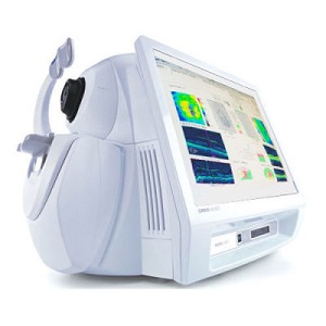
Advancing Smart OCT
for Anterior Segment, Glaucoma and Retina
Anterior Segment Premier Module
PanoMap Wide-Field Display
Smart HD Scans
En face report
Measurement centering with Foveafinder
Visualization at the speed of CIRRUS
Analyzing a single pathology from multiple views provides comprehensive insight and analysis of the clinical situation. How this helps you:
Spot small areas of pathology. Tightly spaced B-scans, (either 30 or 47 μm apart), in the cube ensure that small areas of pathology are imaged. For reference, a human hair is about 40-120 μm in diameter.
Visualize the fovea. Scans that are spaced further apart than in the CIRRUS cube may miss the central fovea.
Fuel for analysis. Millions of data points from the cube are fed into the Zeiss proprietary algorithms for accurate segmentation, reproducible measurements and registration for change analysis.
Take the pressure off the operator. As long as the scan is placed in the vicinity of the fovea or optic nerve, the software automatically centers the measurements after the capture.
See the tissue from different perspectives. View the cube data from all angles, with 3D rendering, OCT fundus images and Advanced Visualization™.
Future ready. Previously captured CIRRUS cubes can be analyzed using new analyses.
Reviews
There are no reviews yet.